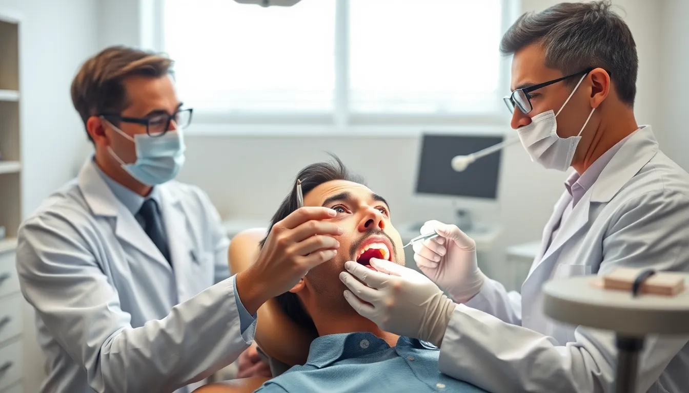

What is furcation in dentistry? If you’ve heard this term from your dentist, it might have sounded concerning—and for good reason. Furcation refers to the area where tooth roots divide, and problems in this region can seriously impact your dental health.
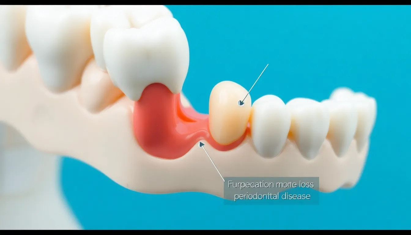
When furcation involvement occurs, the bone between tooth roots begins to deteriorate, creating spaces that trap bacteria and food particles. This condition commonly affects multi-rooted teeth like molars and can be challenging to treat because of the complex anatomy. You’ll want to understand this dental issue thoroughly, as early detection and treatment are crucial for saving affected teeth.
What Is Furcation in Dentistry: An Overview
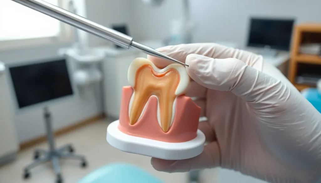
Furcation refers to the anatomical area where a tooth’s roots divide or branch off from the main root trunk. This distinctive feature is present only in multi-rooted teeth, primarily molars and some premolars. Dental professionals categorize furcation as the junction point where the tooth splits into two or more separate roots.
When examining tooth anatomy, you’ll notice that mandibular (lower) molars typically have two roots, while maxillary (upper) molars often have three roots. The spaces between these roots create furcation areas that become vulnerable to periodontal disease when gum health deteriorates.
Furcation involvement occurs when periodontal disease progresses enough to affect the bone between the roots of multi-rooted teeth. These spaces created by bone loss become perfect hiding spots for bacteria and food particles, making them difficult to clean with regular brushing and flossing.
Dr. Todd B. Harris explains, “I’ve seen countless patients who were completely unaware of their furcation involvement until we took radiographs. One patient, Sarah, came in with mild sensitivity in her lower molar. When we examined her, we discovered a Grade II furcation that had been developing silently for years. This underscores why regular dental check-ups are crucial—they help us catch these issues before they become irreversible.”
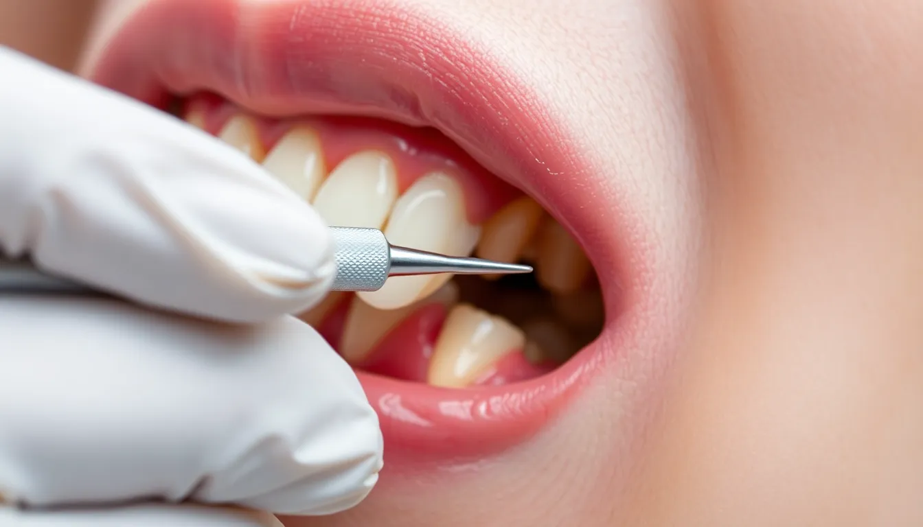
Detecting furcation involvement early is critical for successful treatment outcomes. Dental professionals use specialized probes during clinical examinations to measure the horizontal depth of bone loss in these areas. X-rays complement these examinations by providing visibility into bone loss patterns that aren’t observable during visual inspection.
The complexity of furcation areas creates important challenges for both patients and dental professionals. Their intricate anatomy makes them difficult to access with standard oral hygiene tools, complicating home care efforts. For dentists, these anatomical complexities present unique treatment obstacles that require specialized approaches and techniques.
Anatomy of Tooth Furcation
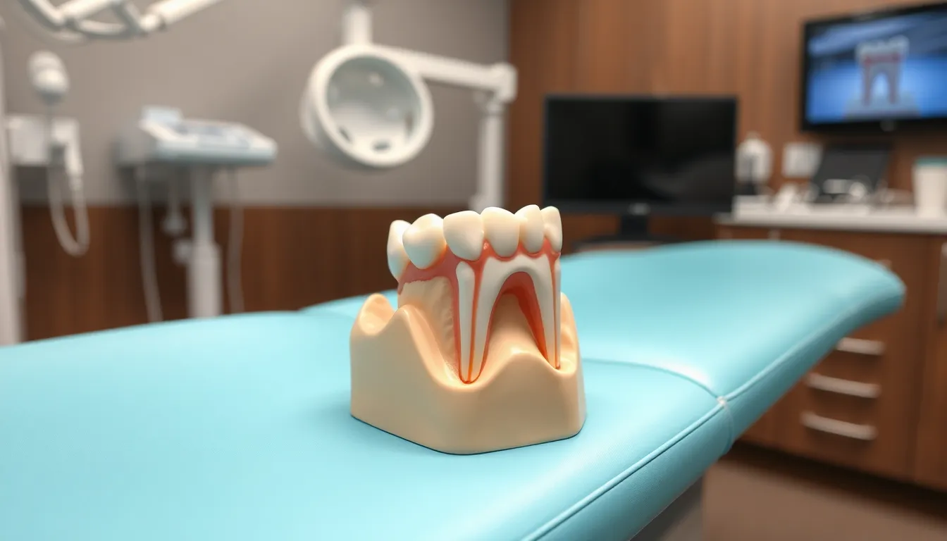
The anatomy of tooth furcation creates a complex structure that’s crucial for dental stability. Understanding these anatomical features helps both dentists and patients appreciate why furcation problems can be challenging to treat.
Types of Furcation Areas
Furcation areas vary depending on the number and arrangement of tooth roots. Bifurcation occurs where two roots diverge and is commonly found in mandibular molars and some premolars. These areas create a natural division that forms a Y-shaped structure below the gumline. Trifurcation, on the other hand, appears where three roots diverge, typically in maxillary molars. The root trunk length directly impacts how quickly furcation involvement can develop during periodontal disease progression. For example, mandibular first molars have shorter root trunks (approximately 3mm buccally and 4mm lingually) compared to maxillary first premolars, which have bifurcations in about 40% of cases with much longer 8mm root trunks.
“I’ve seen many patients surprised to learn about the different root structures in their mouths,” shares Dr. Todd B. Harris. “When I show them models of bifurcated versus trifurcated teeth, they better understand why some areas require more attention during home care.”
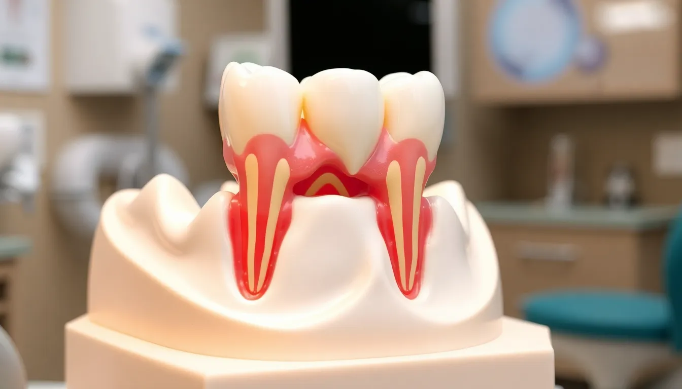
Normal Furcation Structure
A healthy furcation area contains multiple protective elements working together. Intact gingiva covers and shields the furcation entrance, while alveolar bone surrounds both the root trunk and individual roots to provide structural support. Cementum covering the roots adds another layer of protection against bacterial invasion. The interradicular bone fills the space between diverging roots, creating a solid foundation for tooth stability. Root anatomy varies significantly between different teeth – maxillary first molars have root trunks measuring 3-4mm buccally, 4-5mm mesially, and 5-6mm distally. These variations in anatomy explain why furcation problems develop at different rates in different teeth and why treatment approaches must be customized for each exact situation.
The complexity of furcation areas makes them particularly vulnerable when periodontal disease begins. Areas with shorter root trunks typically show furcation involvement earlier in the disease process than those with longer trunks, creating a diagnostic challenge that requires careful probing and radiographic examination.
Furcation Involvement and Classification
Furcation involvement occurs when periodontal disease damages the bone at the junction where tooth roots divide. This condition creates pockets between the roots that trap bacteria and food debris, accelerating bone loss and complicating both professional treatment and home care efforts.
Glickman’s Classification System
Glickman’s classification system categorizes furcation defects into four distinct classes based on severity. Class I represents incipient involvement with minimal bone loss, often detectable only through careful probing. Class II indicates bone destruction extending into the furcation but not completely through it, affecting one-third to two-thirds of the tooth width. Class III describes through-and-through bone loss with an opening large enough for a probe to pass completely through, yet one portion of the furcation roof remains intact. Class IV, the most severe classification, features complete bone destruction with gingival recession that makes the furcation area visible to the naked eye.
“Many patients are shocked when I show them their Class III furcation on a molar they thought was perfectly healthy,” shares Dr. Todd B. Harris. “The challenging part is explaining that what they can’t see is causing important damage beneath the gum line.”
Hamp’s Classification System
Hamp’s classification provides a more measurement-focused approach to furcation defects based on horizontal probing depth. Degree I involves horizontal loss of supporting periodontal tissues up to 3mm. Degree II includes horizontal destruction exceeding 3mm but not completely through the furcation. Degree III represents a “through-and-through” defect where the probe can pass completely from one side to the other.
Dr. Harris notes, “I find Hamp’s classification particularly useful for treatment planning because the exact measurements give patients a clearer understanding of their condition’s severity.” Clinical experience shows that accurate classification directly influences treatment decisions, with early-stage furcation involvement responding better to non-surgical approaches while advanced cases often require surgical intervention or extraction.
Diagnosis of Furcation Defects
Diagnosing furcation defects requires a combination of clinical examination and imaging techniques. Early detection is crucial for determining appropriate treatment options and preventing further progression of periodontal disease in these vulnerable areas.
Clinical Examination Techniques
Clinical probing serves as the primary method for identifying furcation involvement. Your dentist inserts a specialized periodontal probe into the furcation area to measure the depth of attachment loss and assess the degree of involvement. The classification system helps categorize the severity based on how far the probe penetrates horizontally through the furcation. Class I represents early-stage involvement with slight bone loss where the furcation isn’t fully exposed. Class II indicates partial bone loss with a cul-de-sac formation that allows probe entry but not complete passage. Class III signifies through-and-through bone loss where the probe passes completely through the furcation area. Dr. Harris notes, “Many patients are shocked when I demonstrate how my probe can pass completely through what appears to be a solid tooth structure, revealing the extent of their furcation involvement.”
Causes of Furcation Involvement
Furcation involvement develops when the bone supporting multi-rooted teeth deteriorates at the point where roots divide. Several factors contribute to this condition, with periodontal disease being the primary culprit that leads to bone loss in these vulnerable areas.
Periodontal Disease
Periodontal disease stands as the most important cause of furcation involvement. As inflammation progresses through the gum tissues and extends deeper, it gradually destroys the periodontal ligament and alveolar bone supporting the teeth, including the critical furcation regions. This destruction exposes the root structure between diverging roots, creating what dentists call furcation invasion. Dr. Todd B. Harris notes, “Many patients are surprised when I diagnose furcation involvement, as they often don’t experience pain until the condition has significantly advanced.” The complex anatomy of these areas makes them particularly difficult to clean once exposed, accelerating the disease process and creating a challenging treatment scenario.
Other Contributing Factors
Congenital enamel defects play a substantial role in predisposing teeth to furcation problems. These developmental abnormalities create irregular tooth surfaces where enamel folds into the furcation area, preventing proper periodontal ligament attachment and creating pathways for bacterial invasion. Anatomical variations in root trunk length directly impact how quickly furcation involvement develops during periodontal disease—teeth with shorter root trunks typically show signs of involvement earlier than those with longer trunks. Trauma from occlusion, where excessive biting forces damage supporting structures, can accelerate bone loss around furcation areas. Dr. Harris recalls, “I recently treated a patient who had perfect oral hygiene but developed furcation involvement due to untreated grinding habits that created excessive force on his molars.” Endodontic complications resulting from pulpal necrosis can also extend to the furcation area, creating additional pathways for infection and complicating treatment approaches.
Treatment Options for Furcation Defects
Treating furcation defects requires a strategic approach based on the severity of bone loss and individual patient factors. Several treatment modalities exist to address these complex periodontal conditions, ranging from conservative non-surgical methods to advanced surgical techniques.
Non-Surgical Approaches
Non-surgical treatments focus primarily on controlling infection and halting the progression of periodontal disease in furcation areas. Scaling and root planing effectively removes plaque and calculus deposits from accessible portions of the furcation, creating a cleaner environment for healing. Antimicrobial therapy may complement these deep cleaning procedures to reduce bacterial load in the affected areas. Many patients experience improvement with these conservative approaches when furcation involvement is caught early. Dr. Harris often emphasizes to his patients that “consistent home care combined with professional maintenance is crucial for managing early furcation defects, though advanced cases typically require more comprehensive intervention due to the complex anatomy that makes thorough cleaning challenging.”
Surgical Interventions
Surgical procedures provide enhanced access to furcation defects, allowing for more effective treatment of moderate to severe cases. Flap surgery exposes the roots completely, enabling thorough removal of granulation tissue and calculus from previously inaccessible areas. Resective techniques such as root amputation or hemisection involve removing the affected root while preserving the remaining healthy portion of the tooth. Tunneling procedures create accessible channels through the furcation, making daily cleaning possible for patients with through-and-through defects. Dr. Harris recalls a patient with a mandibular molar showing Class III furcation involvement who, after flap surgery, maintained the tooth successfully for years by using specialized interdental brushes to clean the exposed furcation area daily.
Prognosis and Maintenance
Furcation involvement significantly complicates the prognosis of affected teeth due to the challenging architecture that makes bacterial elimination difficult. Teeth with advanced furcation defects typically have a poorer long-term outlook compared to those without such involvement. The prognosis directly correlates with the classification grade—Class I defects generally maintain a favorable outcome with proper care, while Class III and IV defects often face a guarded to poor prognosis.
Root trunk length plays a crucial role in determining how quickly furcation areas become exposed during periodontal disease progression. Mandibular first molars have approximately 3 mm buccal and 4 mm lingual root trunk lengths, making them susceptible to earlier furcation involvement. Maxillary first molars feature varying root trunk lengths: 3-4 mm buccal, 4-5 mm mesial, and 5-6 mm distal, creating different vulnerability patterns across the tooth.
“Many patients are surprised when I explain that their tooth with a Class III furcation defect requires more intensive maintenance than their other teeth,” shares Dr. Todd B. Harris. “I recently treated a patient who diligently maintained a mandibular molar with severe furcation involvement for over 12 years through specialized home care techniques and regular 3-month professional cleanings.”
Successful maintenance of teeth with furcation involvement demands a comprehensive approach:
- Professional cleanings at 3-4 month intervals rather than the standard 6-month schedule
- Specialized cleaning tools such as interdental brushes and water flossers to access furcation areas
- Antimicrobial mouth rinses to reduce bacterial load in difficult-to-clean spaces
- Regular radiographic monitoring to assess stability or progression of bone loss
Patients with treated furcation defects require meticulous oral hygiene practices customized to their exact anatomy. Upper first premolars with bifurcation (occurring in approximately 40% of cases) present unique challenges due to their longer 8 mm root trunk length and less accessible location in the arch.
The management of furcation defects represents a partnership between dental professionals and patients. Treatment success depends not only on the initial intervention but also on the consistent execution of maintenance protocols to prevent reinfection of these vulnerable areas.
Conclusion
Understanding furcation involvement is essential for your long-term dental health. This complex condition requires vigilant detection through regular dental check-ups as it often progresses silently without pain until advanced stages.
Your treatment outcomes depend largely on early intervention and classification severity. While Class I defects typically respond well to non-surgical approaches Class III and IV defects present important challenges requiring more aggressive treatment.
Remember that maintaining teeth with furcation involvement demands a partnership between you and your dental team. With specialized home care tools frequent professional cleanings and consistent monitoring you can often preserve affected teeth for years even though these challenging anatomical considerations.
Don’t underestimate the importance of addressing furcation defects promptly—your diligence can make the difference between keeping your natural teeth and facing extraction.
Frequently Asked Questions
What is furcation in dentistry?
Furcation refers to the area where tooth roots divide in multi-rooted teeth like molars and some premolars. Mandibular (lower) molars typically have two roots, while maxillary (upper) molars often have three. This anatomical feature is crucial for dental stability but can become problematic when affected by periodontal disease.
What is furcation involvement?
Furcation involvement occurs when periodontal disease damages the bone between tooth roots, creating spaces that trap bacteria and food particles. This condition creates pockets that are difficult to clean and can lead to progressive bone loss. Many patients are unaware of this condition until it’s detected during dental examinations.
How are furcation defects classified?
Furcation defects are primarily classified using Glickman’s system, which categorizes them into four classes based on severity: Class I (early involvement), Class II (partial bone loss), Class III (through-and-through bone loss), and Class IV (complete bone destruction). Hamp’s classification system is also used, focusing on horizontal probing depth to guide treatment planning.
How are furcation defects diagnosed?
Dentists diagnose furcation involvement through clinical probing using specialized periodontal probes to measure attachment loss and assess involvement degree. Radiographs (X-rays) are essential for visualizing bone loss patterns and the extent of damage. Early detection is crucial for determining appropriate treatment options and preventing disease progression.
What causes furcation involvement?
Periodontal disease is the primary cause of furcation involvement, as inflammation destroys the periodontal ligament and alveolar bone. Contributing factors include congenital enamel defects, anatomical variations in root trunk length, trauma from occlusion, and endodontic complications. Many patients don’t experience pain until the condition is advanced.
What treatment options exist for furcation defects?
Treatment depends on the severity of bone loss. Non-surgical approaches include scaling and root planing with antimicrobial therapy for early defects. Advanced cases may require surgical interventions such as flap surgery, root amputation, or tunneling procedures. Consistent home care combined with professional maintenance is essential for managing all furcation defects.
What is the prognosis for teeth with furcation involvement?
The prognosis varies based on defect classification. Class I defects generally have favorable outcomes with proper care, while Class III and IV defects often have guarded to poor prognoses. Success depends on defect severity, patient compliance with home care, and regular professional maintenance. Early intervention significantly improves long-term tooth retention.
How should patients maintain teeth with furcation involvement?
Maintenance requires more frequent professional cleanings (typically every 3-4 months), specialized cleaning tools like interdental brushes and water flossers, antimicrobial mouth rinses, and regular radiographic monitoring. Success depends on a partnership between dental professionals and patients, with consistent home care being crucial to prevent reinfection.