Ever wondered why your teeth hurt when you bite down, even without cavities? Traumatic occlusion might be the hidden culprit behind your discomfort, affecting millions of Americans who often mistake it for other dental problems.
When your teeth don’t align properly during biting or chewing, the excessive force creates what dentists call traumatic occlusion. This condition can lead to worn enamel, tooth sensitivity, and even bone loss if left untreated. You might notice symptoms like loose teeth, receding gums, or persistent pain that seems to worsen when eating. Understanding the causes and treatments of traumatic occlusion is crucial for protecting your long-term dental health and preventing more serious complications.
What Is Traumatic Dental Occlusion?
Traumatic dental occlusion refers to abnormal force distribution when your teeth come together during biting or chewing. This condition occurs when teeth don’t align properly, creating excessive pressure on certain teeth or areas of your mouth. Dental professionals categorize traumatic occlusion into primary and secondary types, each with distinct characteristics and causes.
Primary traumatic occlusion happens when excessive forces affect teeth with normal supporting structures. Secondary traumatic occlusion develops when normal biting forces damage teeth that already have compromised supporting structures due to periodontal disease or other issues.
Dr. Todd B. Harris notes, “Many patients come to me confused about why their teeth hurt even though having no visible cavities. Often, the culprit is traumatic occlusion that’s been developing slowly over months or years, gradually wearing down their dental health.”
The biomechanics of traumatic occlusion involve repeated stress that exceeds your teeth’s adaptive capacity. Every time you bite with misaligned teeth, the improper force distribution creates microtrauma that accumulates over time. This persistent trauma eventually manifests as noticeable symptoms and potential complications if left untreated.
Understanding traumatic occlusion helps explain why some patients experience persistent dental pain even though maintaining good oral hygiene. The condition’s impact extends beyond individual teeth, potentially affecting your entire masticatory system including your jaw joints, muscles, and surrounding tissues.
Types of Traumatic Occlusion in Teeth

Traumatic occlusion is divided into two distinct categories based on the condition of supporting structures and the nature of occlusal forces. These classifications help dentists determine appropriate treatment approaches and predict outcomes for patients experiencing tooth pain or mobility.
Primary Traumatic Occlusion
Primary traumatic occlusion develops when excessive occlusal forces affect teeth with healthy periodontal attachment apparatus. Your teeth might be structurally sound with normal gum and bone support, but they’re subjected to forces beyond what they can withstand. Common causes include bruxism (teeth grinding), clenching, off-axis loading during chewing, or habits like nail biting and pencil chewing. These excessive forces vary in magnitude, direction, frequency, and duration, creating a pattern of damage over time.
Microscopically, primary traumatic occlusion can lead to hemorrhage within periodontal tissues, cellular necrosis, widening of the periodontal ligament space, bone resorption, and damage to the cementum layer. The good news is that this condition is often reversible if the source of trauma is identified and eliminated early.
Dr. Todd B. Harris recalls a patient who came in complaining of persistent tooth sensitivity in her upper molars even though having excellent oral hygiene. “After examining her dentition, I noticed tell-tale signs of nighttime grinding. Once we addressed this with a custom nightguard, her symptoms resolved within weeks. This perfectly illustrates how primary traumatic occlusion can be effectively managed when caught early.”
Secondary Traumatic Occlusion
Secondary traumatic occlusion occurs when normal or even excessive occlusal forces act on teeth with already compromised periodontal attachment. Your supporting structures are weakened by pre-existing periodontal disease, making even regular biting forces potentially traumatic. The periodontal ligament and surrounding bone have already lost their integrity, leaving teeth vulnerable to further damage from everyday chewing.
Unlike primary traumatic occlusion, the signs of secondary traumatic occlusion—such as increased tooth mobility—persist even after normalizing occlusal forces. This indicates permanent damage to the supporting structures. Treatment typically requires more comprehensive approaches, often including periodontal therapy, splinting to stabilize affected teeth, and ongoing management of the underlying periodontal condition.
A distinctive feature of secondary traumatic occlusion is that the problem isn’t necessarily the force itself but rather the compromised foundation that can’t withstand normal functional loads. Patients with this condition frequently report progressive tooth loosening and discomfort that worsens over time as periodontal breakdown continues.
Signs and Symptoms of Traumatic Occlusion
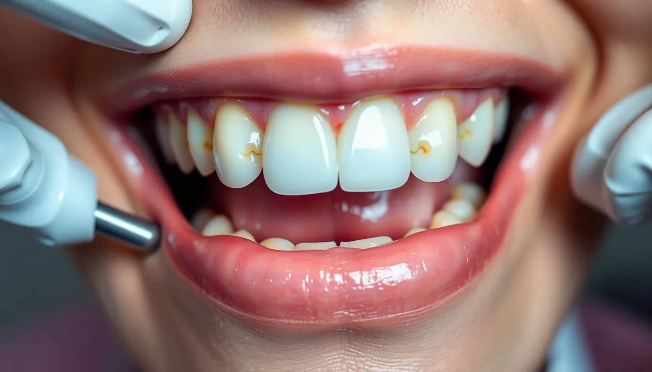
Traumatic occlusion presents with distinctive signs and symptoms that indicate excessive force on your teeth during biting or chewing. These indicators often develop gradually and may appear alongside other dental issues, making proper diagnosis essential for effective treatment.
Clinical Manifestations
Tooth mobility is one of the most common clinical signs of traumatic occlusion, with teeth becoming noticeably loose due to damage to the periodontal ligament and supporting bone. Patients experiencing fremitus—a detectable vibration or movement of teeth during functional contact—often report discomfort when chewing certain foods. Tooth migration occurs as affected teeth shift from their normal positions, creating additional bite problems and aesthetic concerns. Dr. Harris recalls a patient who complained about a front tooth that had gradually shifted forward over several months, creating a visible gap that hadn’t existed before—a classic case of tooth migration due to traumatic occlusion.
Pain and tenderness upon biting or chewing are telltale indicators, with many patients describing the sensation as a sharp discomfort when teeth come together in certain positions. Observable wear facets appear on the occlusal surfaces of affected teeth, particularly on the cusps of molars and premolars, resulting in reduced tooth height and important enamel loss over time. Gingival inflammation often accompanies these symptoms due to trauma and resulting periodontal damage, creating a complex clinical picture that requires comprehensive evaluation.
Radiographic Findings
X-rays reveal distinctive patterns in cases of traumatic occlusion, with widening of the periodontal ligament (PDL) space serving as the primary radiographic indicator. This widening reflects the body’s response to excessive forces as the supporting structures attempt to adapt and absorb the pressure. Thickening of the cervical margin of the alveolar bone appears in many cases, representing another adaptive response to abnormal occlusal loads.
Bone resorption around affected teeth becomes visible on radiographs in more advanced cases, indicating progressive damage to the supporting structures. Loss of cementum—the protective outer layer of the tooth root—may occur, sometimes progressing to necrosis in severe, untreated cases. Dr. Harris emphasizes that these radiographic changes don’t appear overnight but develop gradually as the periodontium responds to ongoing abnormal forces, making regular dental check-ups crucial for early detection. A patient who had ignored minor tooth sensitivity for years was shocked when radiographs revealed important bone loss around several posterior teeth—damage that had occurred silently while only causing minimal symptoms.
Common Causes of Traumatic Dental Occlusion
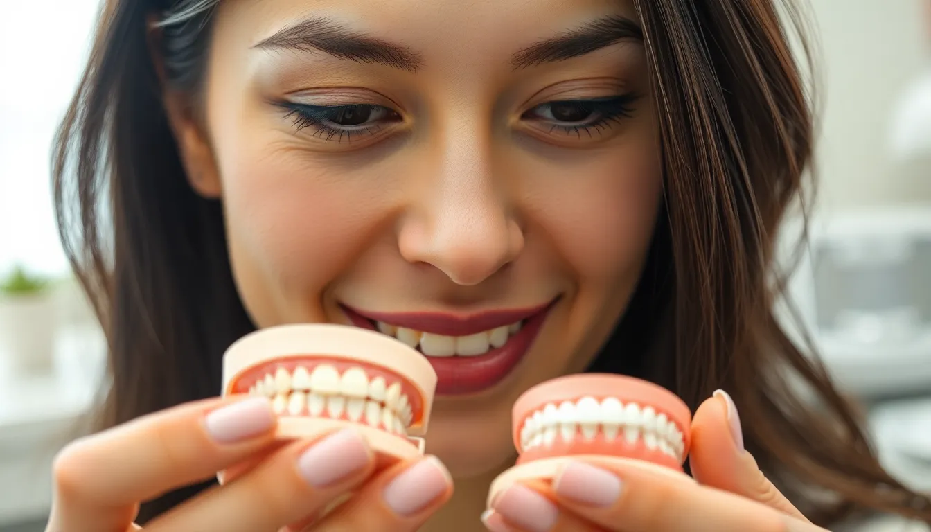
Traumatic dental occlusion stems from exact factors that create excessive or abnormal force during tooth contact. Understanding these causes helps identify why you might experience pain or discomfort when biting down, even without visible dental problems.
Malocclusion Issues
Malocclusion represents a primary cause of traumatic occlusion, occurring when teeth don’t align properly during biting or chewing. Improper tooth alignment creates uneven force distribution that damages teeth and their supporting structures over time. Changes in your bite relationship can result from dental trauma, progressive disease, or even dental treatments that alter the biting surfaces of teeth. Dr. Todd B. Harris notes that many patients are surprised to learn their unexplained tooth pain stems from gradual changes in their bite alignment rather than decay. These alignment issues create stress points where certain teeth bear excessive force, leading to the breakdown of dental structures and persistent discomfort.
Parafunctional Habits
Parafunctional activities involve using your teeth for purposes beyond normal function, creating damaging forces that lead to traumatic occlusion. Bruxism (teeth grinding) and clenching exert tremendous pressure on teeth and supporting structures, often exceeding normal chewing forces by 3-10 times. These habits accelerate tooth wear, creating flat spots on molars and premolars where cusps should normally be prominent. One patient who visited our practice complained of increased tooth sensitivity and jaw pain every morning; after examination, we discovered important wear facets indicating nighttime grinding. Following treatment with a custom nightguard, the patient’s symptoms improved dramatically within weeks. Habits like nail biting, chewing on pens, or using teeth as tools can similarly create abnormal forces that damage periodontal ligaments and compromise tooth stability over time.
Impact of Traumatic Occlusion on Oral Health
Traumatic occlusion creates devastating effects on your entire oral health system when teeth align improperly during biting and chewing. These excessive forces damage not only individual teeth but also their supporting structures, leading to progressive deterioration if left untreated.
Periodontal Damage
Periodontal tissues suffer important damage from traumatic occlusion as abnormal forces create destructive changes in the supporting structures. Your periodontal ligament (PDL) widens and the cervical margin of the alveolar bone thickens in response to these excessive pressures. Clinical signs appear as increasing tooth mobility, migration of teeth from their normal positions, and fremitus—a detectable vibration when touching affected teeth. Bone resorption occurs in advanced cases, potentially compromising tooth stability and leading to tooth loss without proper intervention.
Dr. Todd B. Harris notes, “Many patients come to me confused about why their teeth feel loose even though good oral hygiene. Often, we discover traumatic occlusion has been silently damaging their periodontal structures for months or years before symptoms become obvious.”
Tooth Structure Consequences
Your tooth structure directly experiences the harmful effects of occlusal trauma through several observable changes. Wear facets develop on biting surfaces where improper contact creates excessive friction during chewing. Sensitivity to hot and cold sensations intensifies as protective tooth layers wear away. Pain during chewing or when tapping on teeth indicates internal structural stress that shouldn’t be ignored. Microscopic examination reveals serious damage including hemorrhage, necrosis, and destruction of the cementum—the critical outer layer that anchors your tooth to the periodontal ligament.
One patient’s experience highlights these consequences: “I thought my increasing tooth sensitivity was normal aging until my dentist identified traumatic occlusion as the cause. The wear patterns on my molars showed years of improper biting forces that were slowly destroying my teeth from the outside in.”
Diagnostic signs include tenderness around affected teeth, thermal sensitivity that lingers, mobility when gentle pressure is applied, and visible wear patterns on chewing surfaces. Radiographs reveal widening of the periodontal ligament space and changes in bone density around affected teeth—crucial indicators that help dental professionals assess the severity of your condition.
Diagnosis of Traumatic Occlusion
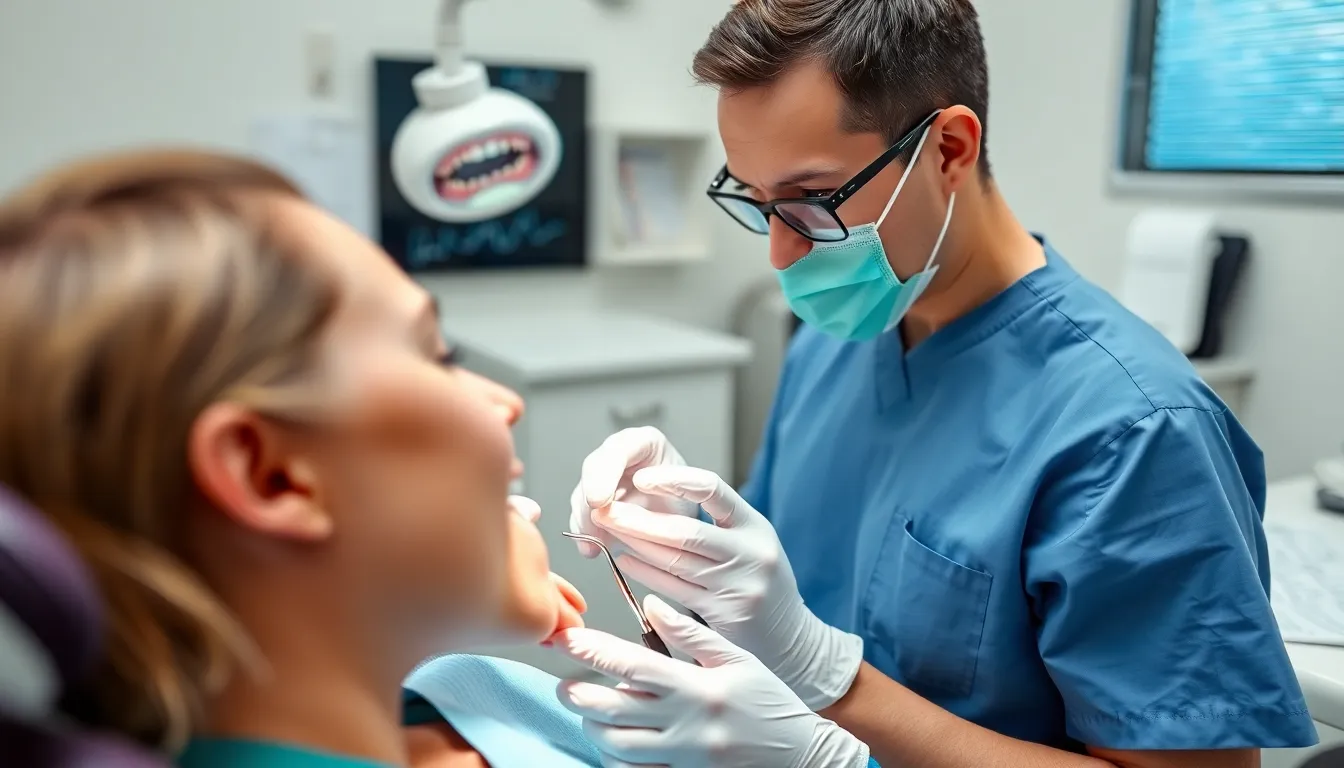
Diagnosing traumatic occlusion requires a comprehensive approach combining clinical examination and radiographic analysis. Since true occlusal trauma confirmation typically requires histological analysis not routinely performed in clinical settings, dentists rely on exact signs and symptoms to identify this condition.
Clinical Examination Techniques
Clinical examination techniques form the foundation for identifying traumatic occlusion in dental patients. Tooth mobility assessment stands as a crucial component of periodontal examination, helping detect looseness caused by occlusal trauma. During your dental appointment, your dentist will gently test each tooth for movement using dental instruments to determine if excessive forces have compromised the supporting structures. Palpation and percussion tests identify areas of tenderness or pain that indicate inflammation or damage resulting from traumatic forces.
Careful observation of wear facets and occlusal prematurity reveals irregular contact points between teeth that create uneven bite forces. Dr. Harris notes, “Many patients are surprised when I show them the wear patterns on their teeth that have developed silently over years of improper biting forces.” The fremitus test, performed by placing a finger on the facial surface of teeth while the patient bites together, detects abnormal tooth movement during occlusion that often indicates traumatic forces. Comprehensive periodontal probing measures attachment loss around teeth, which may be significantly aggravated by occlusal trauma when left untreated.
Advanced Diagnostic Methods
Advanced diagnostic methods provide critical information about the structural impacts of traumatic occlusion that aren’t visible during clinical examination. Dental X-rays and imaging techniques reveal bone density changes, periodontal ligament widening, bone resorption patterns, and alterations in crestal bone levels affected by traumatic forces. These radiographic findings often confirm suspicions raised during clinical examination and help determine the severity of the condition.
Digital occlusal analysis employs computerized tools to measure bite force distribution and occlusal contacts with precise accuracy. This technology helps identify problematic contact points that might be missed during traditional examination. Histological examination, though rarely performed outside research settings, represents the gold standard for confirming occlusal trauma by revealing microscopic evidence of hemorrhage, necrosis, and cementum tears in affected tissues.
Treatment Options for Traumatic Occlusion
Effective treatment for traumatic occlusion focuses on eliminating excessive forces and restoring proper tooth function. Treatment approaches range from conservative methods to surgical interventions, depending on the severity and underlying cause of the condition.
Conservative Approaches
Occlusal adjustment, also known as occlusal equilibration, involves precisely reshaping the biting surfaces of teeth to distribute forces evenly. This technique effectively reduces excessive pressure on exact teeth, preventing further damage to periodontal structures. Many patients experience important relief after these adjustments as the bite becomes more balanced.
Orthodontic therapy offers another conservative solution by realigning teeth and correcting bite issues causing traumatic occlusion. Braces or specialized jaw repositioners can gradually shift teeth into proper positions, eliminating harmful force patterns. Dr. Harris notes, “I’ve seen patients with chronic tooth pain experience complete resolution after proper orthodontic alignment corrected their traumatic occlusion.”
Custom-made night guards provide essential protection for patients with parafunctional habits like grinding or clenching. These appliances create a barrier between upper and lower teeth during sleep, minimizing the impact of excessive forces. A patient of Dr. Harris who suffered from morning headaches and tooth sensitivity for years found immediate relief after using a custom night guard for just two weeks.
Restorative treatments such as dental crowns, inlays, bridges, or partial dentures can rebuild damaged tooth structure while simultaneously improving occlusal relationships. These restorations recreate proper tooth anatomy, allowing for more functional bite patterns and force distribution throughout the dental arch.
Surgical Interventions
Orthognathic surgery becomes necessary in severe cases where traumatic occlusion stems from important jaw misalignment. This procedure repositions the jaws to establish proper occlusion and eliminate destructive forces. Recovery typically takes 6-12 weeks, but the results provide lasting correction of traumatic occlusal patterns.
Tooth repositioning and splinting procedures address teeth displaced by trauma or severe occlusal forces. Digital or forceps repositioning followed by flexible splinting stabilizes affected teeth for 2-4 weeks, allowing periodontal structures to heal. This intervention works best when performed promptly after injury.
Surgical repositioning proves essential for severely intruded teeth (>7 mm) or cases where natural re-eruption hasn’t occurred. Dr. Harris emphasizes, “Intervening within three weeks is critical to prevent ankylosis—a condition where the tooth fuses to surrounding bone, making later treatment nearly impossible.” Patients who receive timely surgical repositioning typically experience better long-term outcomes with reduced risk of tooth loss.
Combining surgical and conservative approaches often yields the best results for complex cases of traumatic occlusion. Your dentist will develop a personalized treatment plan based on your exact condition, addressing both immediate symptoms and underlying causes to restore proper dental function and prevent future complications.
Prevention Strategies
Regular dental check-ups form the foundation of preventing traumatic occlusion. Dentists can identify early signs of occlusal trauma during routine examinations, allowing for timely intervention before important damage occurs. Many patients, like Sarah who visited Dr. Todd B. Harris with minor tooth sensitivity, discover that early detection helped prevent the progression to more severe symptoms.
Breaking harmful oral habits significantly reduces your risk of developing traumatic occlusion. Behaviors such as teeth grinding, nail biting, and thumb sucking contribute to teeth misalignment and create abnormal forces on your dental structures. Eliminating these habits protects your teeth from unnecessary stress and preserves proper alignment.
Protective devices offer effective protection against excessive occlusal forces. Night guards, splints, and similar appliances create a barrier between your upper and lower teeth, particularly benefiting patients prone to bruxism or clenching. Dr. Harris notes, “Custom-fitted night guards have prevented countless cases of severe occlusal trauma among my patients who grind their teeth during sleep.”
Orthodontic and prosthetic treatments correct underlying alignment issues that cause traumatic occlusion. These interventions help achieve balanced bite forces across all teeth, eliminating stress points that lead to damage. Properly aligned teeth function harmoniously during chewing and biting activities, reducing the risk of trauma to individual teeth or exact areas.
Occlusal adjustment therapy involves selective grinding of tooth surfaces to balance contact points. This procedure redistributes biting forces more evenly across your dental arch, though patients should understand that research on its long-term effectiveness continues to evolve. Your dentist can determine if this approach suits your exact condition.
Managing dentinal hypersensitivity early prevents further complications related to traumatic occlusion. Treatments like fluoride varnish or desensitizing resins address tooth sensitivity while protecting vulnerable areas from additional damage. These minimally invasive options provide comfort while supporting the health of your periodontal structures.
Combining these preventive approaches creates a comprehensive strategy against traumatic occlusion. Each measure addresses different aspects of the condition, from behavioral factors to structural issues, working together to maintain optimal oral health and function.
Conclusion
Taking action against traumatic occlusion starts with recognizing its signs and seeking professional help. Don’t ignore tooth sensitivity or discomfort when chewing as these could indicate underlying occlusal issues.
Your dentist can provide personalized treatment options from simple occlusal adjustments to custom night guards or more advanced interventions when necessary. Early detection through regular dental visits remains your best defense against permanent damage.
By addressing harmful habits like teeth grinding and implementing protective measures you’ll not only alleviate current symptoms but also protect your dental health for years to come. Remember that healthy occlusion is fundamental to your overall oral health and quality of life.
Frequently Asked Questions
What is traumatic occlusion?
Traumatic occlusion is a dental condition where abnormal force distribution occurs when teeth come together during biting or chewing. It can cause tooth pain even without cavities present. This improper alignment leads to excessive force on teeth, resulting in symptoms like worn enamel, sensitivity, loose teeth, and persistent pain.
What’s the difference between primary and secondary traumatic occlusion?
Primary traumatic occlusion occurs when excessive forces affect teeth with normal supporting structures, often from habits like teeth grinding. Secondary traumatic occlusion happens when normal biting forces damage teeth with already compromised supporting structures, typically due to periodontal disease. Secondary cases are generally more complex to treat.
What are the common symptoms of traumatic occlusion?
Common symptoms include tooth mobility, discomfort while chewing, tooth migration, visible wear facets on teeth, sensitivity to temperature, receding gums, and persistent pain. These symptoms typically develop gradually over time, which is why many patients don’t notice the condition until significant damage has occurred.
What causes traumatic dental occlusion?
The main causes are malocclusion (improper alignment of teeth) and parafunctional habits like bruxism (teeth grinding) and clenching. Malocclusion leads to uneven force distribution, while parafunctional habits exert excessive pressure on teeth that far exceeds normal chewing forces. Dental trauma, progressive disease, or certain dental treatments can also contribute.
How is traumatic occlusion diagnosed?
Diagnosis involves a comprehensive approach combining clinical examination and radiographic analysis. Dentists assess tooth mobility, perform palpation and percussion tests, and may use the fremitus test to identify occlusal trauma signs. Dental X-rays and digital occlusal analysis provide insights into structural impacts, revealing changes in bone density and occlusal contacts.
What treatment options are available for traumatic occlusion?
Treatment focuses on eliminating excessive forces and restoring proper tooth function. Options range from conservative methods (occlusal adjustment, orthodontic therapy, custom night guards) to surgical interventions (orthognathic surgery, tooth repositioning) for severe cases. Treatment plans are personalized based on the individual’s condition and may combine multiple approaches.
How can I prevent traumatic occlusion?
Prevention includes regular dental check-ups for early detection, breaking harmful habits like teeth grinding and nail biting, using protective devices like night guards for bruxism, seeking orthodontic treatment for alignment issues, and managing dentinal hypersensitivity early. Occlusal adjustment therapy may also help balance biting forces.
Can traumatic occlusion lead to tooth loss?
Yes, if left untreated, traumatic occlusion can eventually lead to tooth loss. The excessive forces damage periodontal tissues, causing widening of the periodontal ligament and changes in the alveolar bone. This results in increasing tooth mobility and, potentially, tooth loss over time.
Is traumatic occlusion painful?
Not always initially. While some patients experience discomfort during chewing or temperature sensitivity, significant damage can occur silently over time without noticeable symptoms. As the condition progresses, symptoms typically include increasing discomfort, sensitivity, and persistent pain.
Can a night guard help with traumatic occlusion?
Yes, custom-made night guards are an effective conservative treatment for traumatic occlusion, especially when caused by bruxism (teeth grinding). They help distribute bite forces evenly and protect teeth from excessive pressure during sleep, preventing further damage and allowing tissues to heal.

